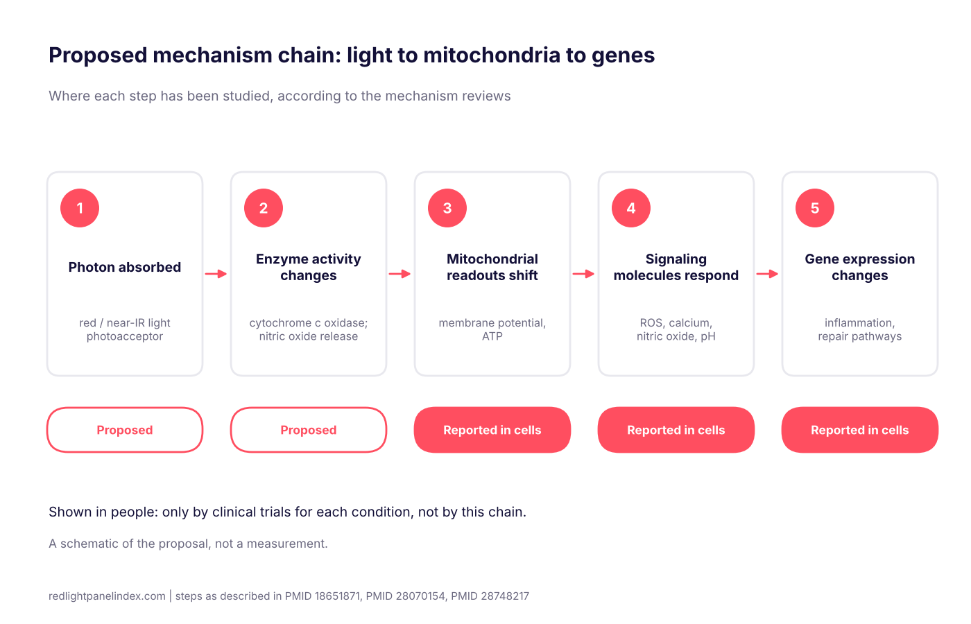
Mitochondria are the compartments inside cells that turn food energy into ATP, the molecule cells spend to do work, and the leading explanation for red light therapy is that red and near-infrared light nudges that process. Researchers describe a chain of steps from photon absorption to changes in cell signaling and gene expression; many steps have been shown in cultured cells, while the link to results in people rests on separate clinical trials.
Choosing a panel? Our ranking of the best red light therapy panels is computed from published, method-labeled specs across 188 devices. For a mechanism like this one, the spec that matters is how much light of an absorbable wavelength reaches the skin, and how that figure was measured.
What mitochondria and ATP do
Almost every cell contains mitochondria. Along their inner membrane sits a set of protein complexes that pass electrons along a chain, pump protons across the membrane, and let an enzyme called ATP synthase use the resulting gradient to make ATP. The membrane potential, the electrical difference across that membrane, is a readout researchers use to gauge how active this system is.
This is ordinary biochemistry taught in every cell biology course. The red light question is narrower: can light of certain colors change how this system behaves, and if so, does that matter for health?
The proposed chain, step by step
The mechanism reviews describe a sequence. The site's photobiomodulation overview summarizes it from the review by de Freitas and Hamblin (PMID 28070154), and Karu's earlier review covers the cell-signaling side (PMID 18651871). Put together, the proposal runs like this:
- Absorption. Red or near-infrared photons are absorbed by a light-sensitive molecule, the photoacceptor. The most discussed candidate is cytochrome c oxidase, the last enzyme in the mitochondrial chain.
- Change in the chain. Absorption is proposed to alter the enzyme's activity, in one popular version by releasing nitric oxide that had been slowing it down; our page on nitric oxide and the blood-flow hypothesis covers how far that idea is supported.
- Mitochondrial readouts shift. Researchers have measured changes after irradiation in mitochondrial membrane potential, ATP, and related signals.
- Signaling molecules respond. Karu's review lists reactive oxygen species, calcium, nitric oxide and intracellular pH among the elements that change in irradiated cells (PMID 18651871). Reactive oxygen species here means the small, short-lived signaling amounts cells use as messages, not the damaging excess associated with oxidative stress.
- Gene expression changes. Signals reach the nucleus and shift which genes are switched on, with proposed effects on processes such as inflammation, cell migration and repair.
A second proposed route involves light-sensitive ion channels that admit calcium into the cell, which the de Freitas and Hamblin review treats alongside the mitochondrial route (PMID 28070154). So the mechanism is not a single pathway, and the reviews present it as a set of proposals.
What has been shown, and where
It helps to separate three levels of evidence.
Shown in cells. Irradiating cultured cells with red and near-infrared light has been reported to change mitochondrial readouts, and the reviews above collect that work. Cells in a dish are controlled systems: known light dose, no skin in the way, often cells that were stressed on purpose.
Proposed for tissue. The step from cells to tissue assumes enough light arrives at the right depth. That depends on penetration depth, which comes mostly from tissue-optics modeling. Hamblin's review of anti-inflammatory effects describes the light-absorbing targets as cytochrome c oxidase and calcium ion channels and reports that downstream effects include more ATP, a brief burst of reactive oxygen species and altered calcium levels (PMID 28748217). It also notes that low doses stimulate while high doses inhibit.
Shown in people only by clinical trials. A cell-level mechanism, however well supported, does not by itself show that a home panel helps a given condition. That question belongs to controlled trials, which is why the site grades each condition separately on its evidence hubs and teaches how to read sham-controlled trials. For example, the muscle recovery evidence page is graded on trial results, not on mitochondrial theory.
Why "more ATP" is not the whole story
Plain-language marketing often compresses all of this into "red light recharges your cells' batteries." That sentence skips several caveats:
- ATP is tightly regulated. Cells do not simply pile it up; what changes in the studies is activity and signaling, and the reviews stress downstream signaling as much as ATP itself (PMID 18651871).
- Different cell types respond differently, and some reported effects are in stressed or damaged cells, not healthy ones.
- The effect depends on dose, and the dose-response is described as biphasic, as covered in the page on the biphasic dose response.
- Several mechanisms are proposed at once, and which dominates may depend on wavelength, tissue and condition.
Key terms in plain language
- Photoacceptor: a molecule that absorbs light and is changed by it. Cytochrome c oxidase is the leading candidate, and ion channels are another.
- Membrane potential: the electrical difference across the inner mitochondrial membrane, used as a gauge of mitochondrial activity.
- Reactive oxygen species: small oxygen-containing molecules that cells use as signals at low levels; the reviews describe a brief burst after light exposure (PMID 28748217).
- Biphasic response: low doses stimulate and high doses inhibit, so more light is not automatically better.
- Downstream signaling: the cascade of messages that carries the effect from the mitochondria to the rest of the cell, including the nucleus.
What wavelengths fit the mechanism
The proposal is about absorption, so it applies to wavelengths the photoacceptors can take up. The mechanism literature centers on red and near-infrared light, and the bands home panels use are laid out in the wavelength guide. The two most common are 660nm red and 850nm near-infrared. The enzyme story does not by itself say which is better for a given goal; the difference in practice is how far light travels in tissue, not a different target in the cell.
A worked example: reading a mechanism claim
Suppose a product page says its panel "boosts mitochondrial ATP." Three checks turn that into something testable:
- Wavelength. Are the panel's peaks in the red and near-infrared range the mechanism literature discusses? A panel's published wavelengths answer this.
- Delivered light. How much irradiance reaches the skin at your distance, and how was it measured? The irradiance explainer shows why a spectrometer reading and a solar meter reading of the same panel can differ.
- Claimed outcome. Does the page stop at the mechanism, or does it claim a result? A mechanism is a reason a result might happen, not evidence that it did.
If a claim satisfies only the first check, it tells you the light is a plausible color and nothing about whether the panel works for you.
What this means for a home panel
For someone choosing or using a panel, the practical points are modest:
- Pick a panel whose published wavelengths fall in the red and near-infrared bands the research addresses.
- Compare irradiance only when the measurement method is stated.
- Keep dose moderate instead of chasing the highest number; the dose calculator turns distance and time into a dose so you can see where a session lands.
- Judge any health claim by the trials for that condition, not by the mitochondrial story.
- Follow general safety guidance, including eye protection and the points on the safety and side effects page.
The mitochondrial mechanism is a good reason to take photobiomodulation seriously as a research field. It is a poor reason to believe any single product claim, and the rest of the answer has to come from the trials.