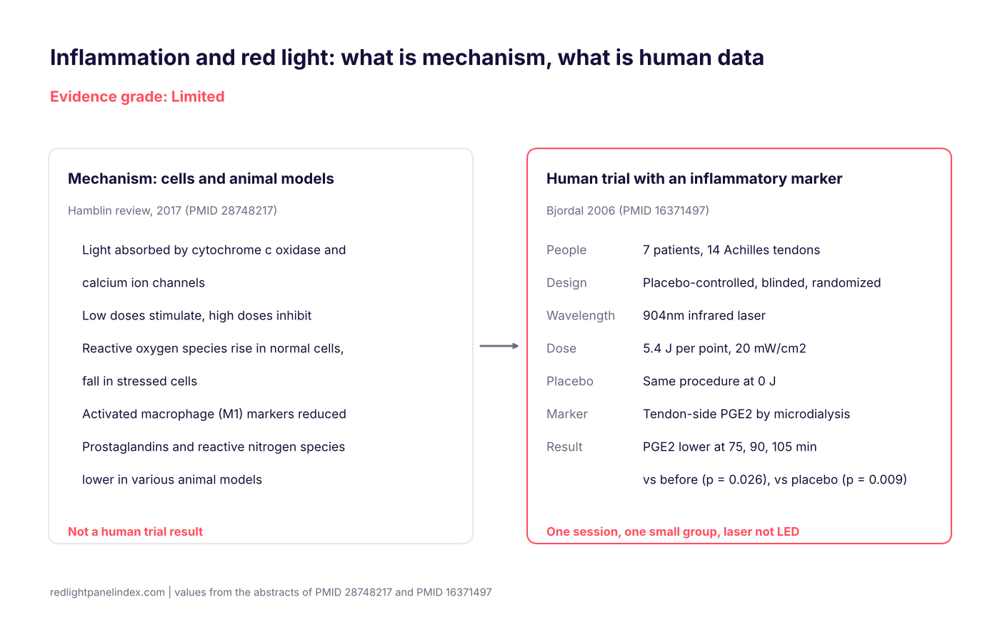
The evidence grade for red and near-infrared light and inflammation is Limited: the mechanism work is extensive but done in cells and animals, and the only human trial in this index's sources that measured an inflammatory marker enrolled seven people and used a 904nm laser, not an LED panel (PMID 16371497). That does not mean light has no effect on inflammation; it means the step from a plausible pathway to a measured human result at panel doses has been tested only once, in a small group, with a device a home panel is not.
Choosing a panel? Our ranking of the best red light therapy panels is computed from published, method-labeled specs across 188 devices. For inflammation questions, the spec that matters is method-labeled irradiance, because the one human trial states its dose and a panel owner needs to know what their own device delivers at distance.
Why the grade is Limited
This site's grading scheme reserves Strong for more than one controlled trial pointing the same way at doses a panel can match. Inflammation as a general claim does not have that. What exists is two different kinds of evidence that should not be added together: a large body of mechanism research, and a very small number of human measurements. Mechanism work explains why an effect might exist. It cannot establish that one does in a person using a home device, and the page that follows keeps the two apart.
"Inflammation" is also not one condition. It is a process inside many conditions, from a sore tendon to arthritis to a skin flare. A marker falling in one tissue after one laser session is not a statement about a whole-body inflammatory state, and nothing in the sources reviewed here supports reading it that way.
The mechanism side: cells and animal models
A 2017 review of the anti-inflammatory effects of photobiomodulation (PMID 28748217) collects the proposed pathways. According to its abstract: Our study review of that paper goes through it section by section.
- Where light is absorbed. The review identifies mitochondrial cytochrome c oxidase and calcium ion channels as the main light-absorbing targets. The cytochrome c oxidase explainer and the page on mitochondria and ATP cover that first step.
- Dose matters in both directions. Low light doses are described as stimulating and high doses as inhibiting, the pattern covered on the biphasic dose response page.
- Oxidative stress. The review describes reactive oxygen species rising in normal cells and falling in stressed cells or disease models. See reactive oxygen species.
- Signaling. It reports that the inflammatory transcription factor NF-kB can be activated in quiescent cells while inflammatory markers fall in activated inflammatory cells.
- Immune cells and mediators. It reports reduced markers of the M1 phenotype in activated macrophages, and lower reactive nitrogen species and prostaglandins in various animal models.
- Where effects were reported. The abstract lists the brain, abdominal fat, wounds, lungs and spinal cord among the tissues in which anti-inflammatory effects have been described.
This is a review, not a trial. It synthesizes cell and animal work and cannot tell you how many minutes on a panel would change anything in a person. The abstract above is the whole of what this page draws from it.
The human side: one small trial with a marker
The Achilles trial (PMID 16371497) is the one human study in this index's sources that put an inflammatory chemical, not only a pain score, at the center. The full study review covers it in detail. In summary:
- Population. Seven patients with bilateral Achilles tendinitis, 14 tendons, with symptoms aggravated by pain-inducing activity immediately before the study. All 14 tendons showed increased blood flow on Doppler ultrasound.
- Device and dose. Infrared laser at 904nm, 5.4 J per point, 20 mW/cm2, against a placebo application of 0 J, in random blinded order.
- Marker. Prostaglandin E2 in the tissue around the tendon, sampled by microdialysis.
- Reported results. PGE2 was significantly lower 75, 90 and 105 minutes after active laser than before treatment (p = 0.026) and than after placebo (p = 0.009). Pressure pain threshold rose significantly after active laser compared with placebo (p = 0.012).
The authors concluded that laser at 5.4 J per point can reduce inflammation and pain in activated Achilles tendinitis. As a reading of what happened in those 14 tendons, that is a fair summary. As a basis for grading inflammation generally, it is thin: seven people, a single treatment, a laser, and a result expressed as p values rather than sizes of change.
What the two sides do not connect
The mechanism review describes a pathway, and the trial found a marker moving in a direction consistent with it. That consistency is worth noting. It is not a test of the pathway. The trial did not measure the cell-level steps the review describes, and the review's animal findings were not tested in the trial's patients.
Other gaps matter more for a panel owner:
- Device. The trial used a 904nm infrared laser applied at points. A panel delivers a different beam over a larger area, typically at 660 and 850nm. The difference between the two device types is covered on the LED versus laser page.
- Dose unit. The trial reports joules per point, while panel specifications use irradiance in mW/cm2. The two cannot be converted without a spot size the abstract does not give.
- Duration. The marker was sampled for roughly an hour and three quarters after one treatment. Nothing in these sources shows how long a change lasts or what repeated sessions do.
What was not studied
None of the sources reviewed here tested, in people:
- a home LED panel at any distance or session length with an inflammatory marker as the outcome;
- blood markers such as C-reactive protein or interleukins after panel use;
- whole-body or systemic inflammation, as distinct from one tendon;
- long-term use, or whether lower prostaglandins translate into better recovery.
Condition pages elsewhere on the site cover trials with clinical outcomes in specific tissues, including tendinopathy and joint pain. Those rest on pain and function measures, which are a different kind of evidence from a tissue marker.
If you use a panel anyway
Nothing here is a reason to avoid red light; it is a reason to keep expectations tied to what was measured. A practical approach:
- Use a method-labeled irradiance figure from your panel's database page and the dose calculator to hold distance and session length constant, so you can tell over several weeks whether anything changes for you.
- Do not escalate the dose. The mechanism literature itself describes a biphasic response in which high doses inhibit. More minutes is not a safe default.
- Match the wavelength to the target. Red and near-infrared reach different depths; the wavelength guide explains the choice.
- Track one outcome you can observe, such as a pain score or range of motion, not an inflammation claim you cannot check.
Cautions
A painful, swollen, hot or red joint or tendon, or swelling after an injury, can have causes that need a clinician and should not be handled with a light panel. Light therapy has not been shown to replace medical treatment of an inflammatory disease. The general safety and side effects page covers eye protection and conditions that call for caution. Products described as treating inflammation are making a claim this evidence does not support at the consumer level.