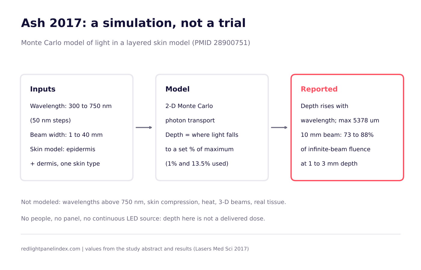
A 2017 paper in Lasers in Medical Science used a computer simulation of a layered skin model and reported that modeled penetration depth increased with wavelength, reaching a maximum of 5378 micrometers (about 5.4 mm), and that widening a beam beyond roughly 10 mm added little extra depth (PMID 28900751). It is a modeling study, not a trial: nobody was treated, no tissue was measured, and the simulation covered visible wavelengths, not the near-infrared bands most home panels use.
Choosing a panel? Our ranking of the best red light therapy panels is computed from published, method-labeled specs across 188 devices. For penetration, the spec that matters is which wavelengths a panel publishes with a stated irradiance, because this simulation says wavelength drives depth far more than spot size does.
What the study asked
The authors, Ash and colleagues, started from a gap they describe plainly: the penetration depth of ultraviolet, visible and infrared light in biological tissue has not been well measured. They were mainly interested in the safety and effectiveness of intense pulsed light and laser sources used in clinics, and they asked how three things change the amount of light energy found at depth: the wavelength, the geometry of the illuminated spot, and skin tone.
For readers of a panel site, the useful part is the first two. If depth is mostly a wavelength question, the spec sheet's wavelength list matters. If spot size barely matters past a point, a larger panel is a coverage choice rather than a depth choice. The site's broader explainer on how deep red and near-infrared light penetrates draws on this paper alongside measured transmission data; this review looks only at the paper itself.
Who was studied
No one. The study is computational, so there is no sample size, no population and no clinical outcome. The "subject" is a mathematical skin model built from layers: an epidermis and a dermis, with optical properties such as refractive index and scattering anisotropy assigned to each layer. Skin tone entered in a limited way. The authors report that skin types from different backgrounds were used to calibrate the wider model against thermography measurements taken after intense pulsed light exposures, while the reported depth simulations used a single fair skin type.
This matters for interpretation. A result from one modeled skin type is not a statement about everyone, and a simulated layer stack is a simplification of real tissue, which has blood vessels, hair follicles, fat, and variation from one body site to the next.
Device and parameters, as stated
The method was a custom Monte Carlo photon transport program, which follows a very large number of simulated photons as they scatter and get absorbed, on a two-dimensional grid. The paper states these inputs:
- Wavelengths: 300 to 750 nm, in steps of 50 nm.
- Beam widths: 1 to 40 mm, with a wider default beam used in the wavelength runs.
- Source: the illumination was modeled on intense pulsed light systems, with fluences in the range of 2 to 20 J/cm2 used for calibration.
- Analysis: results were processed in MATLAB.
There is no irradiance in mW/cm2 for a panel, no session time and no continuous-wave LED source in the model. The paper does not model compression of the skin, heat production or the heat-diffusion step, and the reported runs did not use the heat component.
What was measured and what was found
The main output was a depth at which the modeled light intensity falls to a chosen fraction of the maximum deposited energy. The authors report four findings in the abstract and results:
- Depth rises with wavelength. Across the simulated range, longer wavelengths reached deeper, with a maximum modeled penetration of 5378 micrometers at the long end of the range.
- The threshold matters. Using a 1% criterion, maximum penetration with intense pulsed light was about 5 mm. Using a 13.5% criterion, the figure fell to about 0.37 mm. The same simulation produces very different "depths" depending on how much remaining light you count as meaningful.
- Beam width helps up to a point. Penetration rose as the beam widened from 1 mm to about 5 mm, then leveled off near 10 mm. A 10 mm beam delivered 73 to 88% of the fluence an infinitely wide beam would deliver at depths of 1 to 3 mm.
- The skin surface boundary matters. The authors report that absorption changes sharply where the epidermis meets the dermis, which shapes how much energy continues deeper.
Number 2 is the one most worth remembering. When a source says light "penetrates X millimeters," ask which cutoff it used. This paper shows one model can give a result near 5 mm or well under 1 mm depending on the choice, which is why the site treats depth figures from different methods as non-comparable.
Limitations
The authors list several, and a reader should add a few more.
- Two-dimensional model. Real light spreads in three dimensions, and the beam was treated as uniform rather than shaped like a real device's output.
- Relative, not absolute, intensity. Depth is expressed as a percentage of maximum deposited energy, not as an incident intensity, and light that escapes the tissue is excluded, so the authors note that actual penetration would be smaller than their reported depths.
- Limited wavelength range. Only 300 to 750 nm was simulated. Wavelengths above 750 nm, including the 810, 830 and 850 nm bands common in home panels, were not modeled, and finer steps were not explored.
- No compression or heat. Pressing a device against skin can change effective depth, and the paper did not model it. Temperature at depth is unknown.
- No agreed definition. There is no accepted definition of a therapeutic penetration depth, so the 1% and 13.5% cutoffs are conventions, not biological thresholds.
- From the review side: the work is aimed at pulsed light clinics, it is one skin type, and it has no validation against measured transmission through living tissue in this paper.
What it means for a home panel
Three practical readings hold up, and each is modest.
First, the direction is consistent with the idea behind the optical window: within the range simulated, longer wavelengths travel further. That supports reading a panel's wavelength list rather than treating "red light" as one thing. The paper does not, however, test the near-infrared bands, so it cannot tell you how much deeper 850 nm goes than 660 nm. See the wavelength guide for what each band is used for.
Second, spot size is a weaker lever than many assume. A panel's beam angle and size affect how evenly light covers an area, but in this model a beam past about 10 mm added little depth. A larger panel treats more skin; it should not be expected to reach deeper on the strength of this study.
Third, the simulation says nothing about dose. A panel delivers continuous LED light at a stated irradiance for minutes, while the model describes brief intense pulses. To size a session for a real panel, use a measured irradiance with a stated method, such as those explained in the irradiance guide, and run the numbers through the dose calculator. Depth in a simulation is not a delivered dose in tissue.
The honest gap between this paper and a panel is large: a pulsed-light model, visible wavelengths only, one skin type, no measured validation here. It is a useful piece of physics for understanding why wavelength matters, and a poor basis for any claim about how many millimeters a particular panel reaches.